
The ability of fireflies to glow in the dark, delights those who have seen these insects in action. It really seems like a special talent. However a recent issue of National Geographic (March 2015) declared about bioluminescence: “Evolving to make light seems to be relatively easy — it has happened independently in at least 40 different lineages.” (p. 84) Just because we find a special talent in a number of very different creatures, does not mean that the talent was easily developed by chance. National Geographic is not aware that this unusual ability is much more reasonably explained as the choice of God, the creator. There are many examples where we can see the problem for evolution of special talents in very different creatures. And the camera eye is an ideal example.
Everybody knows that our eyes are wonderfully designed. All the parts are special and each is important for vision. The bulging cornea consists of clear material which bends light toward the pupil. The iris consists of a thin circular muscle which acts like a camera diaphragm. The iris expands or contracts the pupil opening in order to control the amount of light entering the eye. Behind the pupil is the lens which focuses light onto the retina (composed of light sensitive cells and nerve cells).
The oval-shaped lens is made up of water soluble proteins, many of which are very large molecules. These proteins are tightly packed together in such a way that they are not only transparent, but they bend the light so that the rays are focused into a sharp point. This provides a clear image. Ideally the lens focuses on the retina (the receiver), but if the focal point is in front of the retina (or behind it) then corrective lenses are required to adjust the focus onto the retina. It is also most important that the proteins in the lens retain their special tightly packed arrangement, otherwise the lens becomes cloudy thereby disturbing vision.
The other particularly important component of the eye is the retina. It consists of certain receiver cells which contain light sensitive pigments called rhodopsins. These are composed of a form of vitamin A and a large protein molecule called opsin. Different precisely shaped opsins are sensitive to specific wavelengths of light. Before the light gets to the rods and cones (in the retina), it passes through the nerve cells which lie on top of the light sensitive cells. Some people suggest that this is backward wiring and not the most efficient arrangement of parts. But what do they know? Others suggest that this arrangement protects the sensitive tissue from too much light. The nerve cells lying on top of the rods and cones, then conduct an electrical impulse (generated by the rods and cones) to the optic nerve and to the brain. The brain, for its part, puts the electrical signals together into images which are communicated to the person’s consciousness.
There are other important components of the camera eye too, like the dark layer lining the inner eyeball, which prevents light rays from scattering inside the eye, and jelly-like material which allows the eye to keep its shape. When we consider the special properties of all these component parts, we have to conclude that the camera eye is indeed a wonderful organ.
Among living creatures there are other eye designs as well. Some single celled animals and even some much larger creatures make do with clusters of pigmented cells. In some many celled animals, these are often associated with nerve cells. Creatures with jointed outside skeletons (exoskeletons) like insects, are famous for their composite eyes. These bulbous structures are made up of many tiny eyes all of which focus on a central point. While these eyes are very good at detecting motion, they probably do not have the same sharp focus as the camera eye.
All creatures with backbones (vertebrates) enjoy camera style eyes. Most of us know that! But what about a jellyfish, octopus and a single celled animal which closely resembles algae that cause toxic red tides in the sea? Do they have camera style eyes too? Yes, yes and yes!
Octopus and squid are perhaps the best known animals without a backbone (invertebrates) which enjoy the benefits of a camera-style eye. Octopi are particularly intelligent, some say as intelligent as a housecat. The term cephalopod means brainy foot and it denotes a subgroup of mollusks which include squid and octopus. The cephalopod camera-type eye includes an iris, circular lens, gel filling the eyeball, pigment cells and photoreceptor cells that send an electrical signal to the optic nerve which is connected to the brain. In the case of the cephalopods, the light sensitive rods and cones are in front of the nerve cells (not behind as in vertebrates). Moreover the crystal proteins in the cephalopod lens act the same way as our lens does, but the proteins are not the same. In that the cephalopods have a body design (plan) which is far different from that of vertebrates, and in that the chemical components of the eye are different, not even mainstream scientists see any kind of evolutionary connection between us and the octopus.
We are all familiar with earthworms. These creatures have a complete digestive tract with a mouth at one end and an anus at the other end. They have strong muscles and a few projecting bristles, but no obvious sense organs although they react strongly to odours and the drying effects of light. This body plan possessed by a group called the annelids, does not seem promising for fancy sense organs. However there are marine annelids called polychaetes (meaning many bristles) which lead more vigorous lifestyles. Among the polychaetes is an obscure group called alciopids. These are slender swimming creatures with conspicuous eyes. They actively pursue and catch prey. Most surprisingly, the eyes of these worms are camera style eyes complete with cornea, lens and retina, and like cephalopods, the wiring of the retina features the light sensitive cells first with the transmitting nerve cells behind. Obviously there is nothing in the body plan of these annelids that is at all similar to vertebrates. So nobody imagines that there is a shared evolutionary history between the two groups.
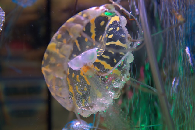
Even the annelids have some small concentrations of nerve cells at the front end of the body. If there is going to be any interpretation of the images detected by the fancy eye, it would be in this “brain”. Jellyfish however have no central nervous tissue (which could function as some sort of brain). These creatures therefore do not look like promising candidates for any benefit from camera-style eyes. Nevertheless box jellyfish indeed possess camera-style eyes. One commentator called attention to the surprising occurrence of this eye design in any creature with a jellyfish body plan: “…. — a box jellyfish or cubomedusa — is equipped with eight surprisingly sophisticated lens eyes of the camera-type, but there is no common brain behind them. In nearly every respect, these lens eyes resemble those of animals such as fish or cephalopods, but the ‘central nervous system’ behind the eyes consists only of a diffuse nerve net accompanied by a marginal nerve ring.” (Rudiger Wehner. Nature 435 (7039) May 12, 2005 p. 157)
Jellyfish mostly drift or swim in the open sea in such a way that their trailing tentacles occasionally encounter suitable prey. Specially designed stinging cells entangle and kill the prey and the tentacles pull the victim to a central opening that serves both as the mouth and anus. However the box jellyfish actively hunt prey in shallow water habitats like mangrove swamps which are full of obstructions like tree roots.
Despite the unexpected nature of the box jellyfish sensory organs, expert Dan Nilsson insists: “All major components of a typical camera-type eye are present: a cornea, a lens, a retina, a pigment layer and an iris.” (Nature 435 (7039) May 12, 2005 p. 202) Not only do the jellyfish eyes have all the appropriate parts, but it transpires that the jellyfish lens produces a very sharp focus. Dr. Nilsson and team declare that such lenses are not only rarely encountered among the variety of animal body plans, but the jellyfish lenses are unique by virtue of special proteins: “From the unique crystalline proteins we know that at least the lenses have evolved independently in box jellyfish. Making good lenses seems to be a demanding task because only a few animal phyla have accomplished it.” (p. 202)
Despite the sharp focus of the jellyfish lens, the retina is positioned too close so that a blurred image results. Nilsson and colleagues however suggest that the eyes are “‘purposely’ underfocused” (p. 202) so that the creature is not confused by too much detail. The lack of brain may also suit the lifestyle of this animal as commentator Rudiger Wehner reports: “… box jellyfish have clearly not had the need to feed the information provided by their total of 24 eyes into a central processing unit, or brain.” (p. 159)
In its body plan the box jellyfish is completely unlike other animals with camera-style eyes which typically possess some sort of central brain. In its body plan a jellyfish exhibits minimal body parts, but in the case of the box jellyfish we also see a sensory organ which follows a precise sophisticated blueprint. The great differences with other creatures of similar eye design mean that no evolutionary relationship is imagined between eye-possessing box jellyfish, polychaete worms, octopi and vertebrates. It was in some other way that they came to possess the fancy eye blueprint.
If camera style eyes in a jellyfish are unexpected, how weird would it be to see the same design in a single-celled animal? A recent article in Nature however communicated the astounding news that there are some single celled protozoans that have a sensory structure “so complex that it was initially mistaken for a multicellular eye.” (523 (7559) July 9, 2015 p. 204) The component parts include a cornea, lens, iris and retina. It is these parts, which, declares Gregory Gavelis and colleagues “so resemble the camera-type eye of some animals that they have been speculated to be homologous [related through evolutionary descent].” (p. 204)
Warnowiid dinoflagellates are very rare and unusual marine organisms. Their cell design is like the algae that cause toxic red tides in oceans. These dense concentrations of algae can kill fish and render shellfish (which consume the algae) poisonous to people. Most dinoflagellates are dark brown and photosynthetic (manufacturing their own food). The warnowiid dinoflagellates however are colourless and need to consume food. Presumably these cells use their ocelloid (eye-like structure) to catch suitable prey.
Commenting on the dinoflagellate study (which came from University of British Columbia), commentators declare: “evolution has stumbled on similar solutions to perceiving light time and time again.” (Nature July 9. 2015 p. 167) It is true that in the course of this survey of creatures with camera-style eyes we have observed that (apart from vertebrates), the possessors are rare specimens from diverse body plans. Obviously there was no line of descent. The creatures are too different to even contemplate such an idea. Mainstream scientists instead contemplate the separate surprising appearance of the same blueprint/design in wildly different organisms by means of evolutionary processes which converge from highly different sources on the same solution to lifestyle problems. However in the cases which we have discussed, the lifestyles are not even remotely similar, so it would be surprising to see similar solutions, especially through chance processes. Alternatively what we see is common design (conscious choice by the Creator) rather than descent with change from a single ancestral population (common descent) or separate spontaneous appearances in diverse creatures. When we see these examples as the work of God, our appreciation of the creation becomes much more profound.
Margaret Helder
October 2015
Subscribe to Dialogue
FEATURED BOOKS AND DVDS

Paperback / $6.00 / 55 Pages
The ability of fireflies to glow in the dark, delights those who have seen these insects in action. It really seems like a special talent. However a recent issue of National Geographic (March 2015) declared about bioluminescence: “Evolving to make light seems to be relatively easy — it has happened independently in at least 40 different lineages.” (p. 84) Just because we find a special talent in a number of very different creatures, does not mean that the talent was easily developed by chance. National Geographic is not aware that this unusual ability is much more reasonably explained as the choice of God, the creator. There are many examples where we can see the problem for evolution of special talents in very different creatures. And the camera eye is an ideal example.
Everybody knows that our eyes are wonderfully designed. All the parts are special and each is important for vision. The bulging cornea consists of clear material which bends light toward the pupil. The iris consists of a thin circular muscle which acts like a camera diaphragm. The iris expands or contracts the pupil opening in order to control the amount of light entering the eye. Behind the pupil is the lens which focuses light onto the retina (composed of light sensitive cells and nerve cells).
The oval-shaped lens is made up of water soluble proteins, many of which are very large molecules. These proteins are tightly packed together in such a way that they are not only transparent, but they bend the light so that the rays are focused into a sharp point. This provides a clear image. Ideally the lens focuses on the retina (the receiver), but if the focal point is in front of the retina (or behind it) then corrective lenses are required to adjust the focus onto the retina. It is also most important that the proteins in the lens retain their special tightly packed arrangement, otherwise the lens becomes cloudy thereby disturbing vision.
The other particularly important component of the eye is the retina. It consists of certain receiver cells which contain light sensitive pigments called rhodopsins. These are composed of a form of vitamin A and a large protein molecule called opsin. Different precisely shaped opsins are sensitive to specific wavelengths of light. Before the light gets to the rods and cones (in the retina), it passes through the nerve cells which lie on top of the light sensitive cells. Some people suggest that this is backward wiring and not the most efficient arrangement of parts. But what do they know? Others suggest that this arrangement protects the sensitive tissue from too much light. The nerve cells lying on top of the rods and cones, then conduct an electrical impulse (generated by the rods and cones) to the optic nerve and to the brain. The brain, for its part, puts the electrical signals together into images which are communicated to the person’s consciousness.
There are other important components of the camera eye too, like the dark layer lining the inner eyeball, which prevents light rays from scattering inside the eye, and jelly-like material which allows the eye to keep its shape. When we consider the special properties of all these component parts, we have to conclude that the camera eye is indeed a wonderful organ.
Among living creatures there are other eye designs as well. Some single celled animals and even some much larger creatures make do with clusters of pigmented cells. In some many celled animals, these are often associated with nerve cells. Creatures with jointed outside skeletons (exoskeletons) like insects, are famous for their composite eyes. These bulbous structures are made up of many tiny eyes all of which focus on a central point. While these eyes are very good at detecting motion, they probably do not have the same sharp focus as the camera eye.
All creatures with backbones (vertebrates) enjoy camera style eyes. Most of us know that! But what about a jellyfish, octopus and a single celled animal which closely resembles algae that cause toxic red tides in the sea? Do they have camera style eyes too? Yes, yes and yes!
Octopus and squid are perhaps the best known animals without a backbone (invertebrates) which enjoy the benefits of a camera-style eye. Octopi are particularly intelligent, some say as intelligent as a housecat. The term cephalopod means brainy foot and it denotes a subgroup of mollusks which include squid and octopus. The cephalopod camera-type eye includes an iris, circular lens, gel filling the eyeball, pigment cells and photoreceptor cells that send an electrical signal to the optic nerve which is connected to the brain. In the case of the cephalopods, the light sensitive rods and cones are in front of the nerve cells (not behind as in vertebrates). Moreover the crystal proteins in the cephalopod lens act the same way as our lens does, but the proteins are not the same. In that the cephalopods have a body design (plan) which is far different from that of vertebrates, and in that the chemical components of the eye are different, not even mainstream scientists see any kind of evolutionary connection between us and the octopus.
We are all familiar with earthworms. These creatures have a complete digestive tract with a mouth at one end and an anus at the other end. They have strong muscles and a few projecting bristles, but no obvious sense organs although they react strongly to odours and the drying effects of light. This body plan possessed by a group called the annelids, does not seem promising for fancy sense organs. However there are marine annelids called polychaetes (meaning many bristles) which lead more vigorous lifestyles. Among the polychaetes is an obscure group called alciopids. These are slender swimming creatures with conspicuous eyes. They actively pursue and catch prey. Most surprisingly, the eyes of these worms are camera style eyes complete with cornea, lens and retina, and like cephalopods, the wiring of the retina features the light sensitive cells first with the transmitting nerve cells behind. Obviously there is nothing in the body plan of these annelids that is at all similar to vertebrates. So nobody imagines that there is a shared evolutionary history between the two groups.
Even the annelids have some small concentrations of nerve cells at the front end of the body. If there is going to be any interpretation of the images detected by the fancy eye, it would be in this “brain”. Jellyfish however have no central nervous tissue (which could function as some sort of brain). These creatures therefore do not look like promising candidates for any benefit from camera-style eyes. Nevertheless box jellyfish indeed possess camera-style eyes. One commentator called attention to the surprising occurrence of this eye design in any creature with a jellyfish body plan: “…. — a box jellyfish or cubomedusa — is equipped with eight surprisingly sophisticated lens eyes of the camera-type, but there is no common brain behind them. In nearly every respect, these lens eyes resemble those of animals such as fish or cephalopods, but the ‘central nervous system’ behind the eyes consists only of a diffuse nerve net accompanied by a marginal nerve ring.” (Rudiger Wehner. Nature 435 (7039) May 12, 2005 p. 157)
Jellyfish mostly drift or swim in the open sea in such a way that their trailing tentacles occasionally encounter suitable prey. Specially designed stinging cells entangle and kill the prey and the tentacles pull the victim to a central opening that serves both as the mouth and anus. However the box jellyfish actively hunt prey in shallow water habitats like mangrove swamps which are full of obstructions like tree roots.
Despite the unexpected nature of the box jellyfish sensory organs, expert Dan Nilsson insists: “All major components of a typical camera-type eye are present: a cornea, a lens, a retina, a pigment layer and an iris.” (Nature 435 (7039) May 12, 2005 p. 202) Not only do the jellyfish eyes have all the appropriate parts, but it transpires that the jellyfish lens produces a very sharp focus. Dr. Nilsson and team declare that such lenses are not only rarely encountered among the variety of animal body plans, but the jellyfish lenses are unique by virtue of special proteins: “From the unique crystalline proteins we know that at least the lenses have evolved independently in box jellyfish. Making good lenses seems to be a demanding task because only a few animal phyla have accomplished it.” (p. 202)
Despite the sharp focus of the jellyfish lens, the retina is positioned too close so that a blurred image results. Nilsson and colleagues however suggest that the eyes are “‘purposely’ underfocused” (p. 202) so that the creature is not confused by too much detail. The lack of brain may also suit the lifestyle of this animal as commentator Rudiger Wehner reports: “… box jellyfish have clearly not had the need to feed the information provided by their total of 24 eyes into a central processing unit, or brain.” (p. 159)
In its body plan the box jellyfish is completely unlike other animals with camera-style eyes which typically possess some sort of central brain. In its body plan a jellyfish exhibits minimal body parts, but in the case of the box jellyfish we also see a sensory organ which follows a precise sophisticated blueprint. The great differences with other creatures of similar eye design mean that no evolutionary relationship is imagined between eye-possessing box jellyfish, polychaete worms, octopi and vertebrates. It was in some other way that they came to possess the fancy eye blueprint.
If camera style eyes in a jellyfish are unexpected, how weird would it be to see the same design in a single-celled animal? A recent article in Nature however communicated the astounding news that there are some single celled protozoans that have a sensory structure “so complex that it was initially mistaken for a multicellular eye.” (523 (7559) July 9, 2015 p. 204) The component parts include a cornea, lens, iris and retina. It is these parts, which, declares Gregory Gavelis and colleagues “so resemble the camera-type eye of some animals that they have been speculated to be homologous [related through evolutionary descent].” (p. 204)
Warnowiid dinoflagellates are very rare and unusual marine organisms. Their cell design is like the algae that cause toxic red tides in oceans. These dense concentrations of algae can kill fish and render shellfish (which consume the algae) poisonous to people. Most dinoflagellates are dark brown and photosynthetic (manufacturing their own food). The warnowiid dinoflagellates however are colourless and need to consume food. Presumably these cells use their ocelloid (eye-like structure) to catch suitable prey.
Commenting on the dinoflagellate study (which came from University of British Columbia), commentators declare: “evolution has stumbled on similar solutions to perceiving light time and time again.” (Nature July 9. 2015 p. 167) It is true that in the course of this survey of creatures with camera-style eyes we have observed that (apart from vertebrates), the possessors are rare specimens from diverse body plans. Obviously there was no line of descent. The creatures are too different to even contemplate such an idea. Mainstream scientists instead contemplate the separate surprising appearance of the same blueprint/design in wildly different organisms by means of evolutionary processes which converge from highly different sources on the same solution to lifestyle problems. However in the cases which we have discussed, the lifestyles are not even remotely similar, so it would be surprising to see similar solutions, especially through chance processes. Alternatively what we see is common design (conscious choice by the Creator) rather than descent with change from a single ancestral population (common descent) or separate spontaneous appearances in diverse creatures. When we see these examples as the work of God, our appreciation of the creation becomes much more profound.
Order Online
Hardcover / $35.00 / 295 Pages / Full colour
The ability of fireflies to glow in the dark, delights those who have seen these insects in action. It really seems like a special talent. However a recent issue of National Geographic (March 2015) declared about bioluminescence: “Evolving to make light seems to be relatively easy — it has happened independently in at least 40 different lineages.” (p. 84) Just because we find a special talent in a number of very different creatures, does not mean that the talent was easily developed by chance. National Geographic is not aware that this unusual ability is much more reasonably explained as the choice of God, the creator. There are many examples where we can see the problem for evolution of special talents in very different creatures. And the camera eye is an ideal example.
Everybody knows that our eyes are wonderfully designed. All the parts are special and each is important for vision. The bulging cornea consists of clear material which bends light toward the pupil. The iris consists of a thin circular muscle which acts like a camera diaphragm. The iris expands or contracts the pupil opening in order to control the amount of light entering the eye. Behind the pupil is the lens which focuses light onto the retina (composed of light sensitive cells and nerve cells).
The oval-shaped lens is made up of water soluble proteins, many of which are very large molecules. These proteins are tightly packed together in such a way that they are not only transparent, but they bend the light so that the rays are focused into a sharp point. This provides a clear image. Ideally the lens focuses on the retina (the receiver), but if the focal point is in front of the retina (or behind it) then corrective lenses are required to adjust the focus onto the retina. It is also most important that the proteins in the lens retain their special tightly packed arrangement, otherwise the lens becomes cloudy thereby disturbing vision.
The other particularly important component of the eye is the retina. It consists of certain receiver cells which contain light sensitive pigments called rhodopsins. These are composed of a form of vitamin A and a large protein molecule called opsin. Different precisely shaped opsins are sensitive to specific wavelengths of light. Before the light gets to the rods and cones (in the retina), it passes through the nerve cells which lie on top of the light sensitive cells. Some people suggest that this is backward wiring and not the most efficient arrangement of parts. But what do they know? Others suggest that this arrangement protects the sensitive tissue from too much light. The nerve cells lying on top of the rods and cones, then conduct an electrical impulse (generated by the rods and cones) to the optic nerve and to the brain. The brain, for its part, puts the electrical signals together into images which are communicated to the person’s consciousness.
There are other important components of the camera eye too, like the dark layer lining the inner eyeball, which prevents light rays from scattering inside the eye, and jelly-like material which allows the eye to keep its shape. When we consider the special properties of all these component parts, we have to conclude that the camera eye is indeed a wonderful organ.
Among living creatures there are other eye designs as well. Some single celled animals and even some much larger creatures make do with clusters of pigmented cells. In some many celled animals, these are often associated with nerve cells. Creatures with jointed outside skeletons (exoskeletons) like insects, are famous for their composite eyes. These bulbous structures are made up of many tiny eyes all of which focus on a central point. While these eyes are very good at detecting motion, they probably do not have the same sharp focus as the camera eye.
All creatures with backbones (vertebrates) enjoy camera style eyes. Most of us know that! But what about a jellyfish, octopus and a single celled animal which closely resembles algae that cause toxic red tides in the sea? Do they have camera style eyes too? Yes, yes and yes!
Octopus and squid are perhaps the best known animals without a backbone (invertebrates) which enjoy the benefits of a camera-style eye. Octopi are particularly intelligent, some say as intelligent as a housecat. The term cephalopod means brainy foot and it denotes a subgroup of mollusks which include squid and octopus. The cephalopod camera-type eye includes an iris, circular lens, gel filling the eyeball, pigment cells and photoreceptor cells that send an electrical signal to the optic nerve which is connected to the brain. In the case of the cephalopods, the light sensitive rods and cones are in front of the nerve cells (not behind as in vertebrates). Moreover the crystal proteins in the cephalopod lens act the same way as our lens does, but the proteins are not the same. In that the cephalopods have a body design (plan) which is far different from that of vertebrates, and in that the chemical components of the eye are different, not even mainstream scientists see any kind of evolutionary connection between us and the octopus.
We are all familiar with earthworms. These creatures have a complete digestive tract with a mouth at one end and an anus at the other end. They have strong muscles and a few projecting bristles, but no obvious sense organs although they react strongly to odours and the drying effects of light. This body plan possessed by a group called the annelids, does not seem promising for fancy sense organs. However there are marine annelids called polychaetes (meaning many bristles) which lead more vigorous lifestyles. Among the polychaetes is an obscure group called alciopids. These are slender swimming creatures with conspicuous eyes. They actively pursue and catch prey. Most surprisingly, the eyes of these worms are camera style eyes complete with cornea, lens and retina, and like cephalopods, the wiring of the retina features the light sensitive cells first with the transmitting nerve cells behind. Obviously there is nothing in the body plan of these annelids that is at all similar to vertebrates. So nobody imagines that there is a shared evolutionary history between the two groups.
Even the annelids have some small concentrations of nerve cells at the front end of the body. If there is going to be any interpretation of the images detected by the fancy eye, it would be in this “brain”. Jellyfish however have no central nervous tissue (which could function as some sort of brain). These creatures therefore do not look like promising candidates for any benefit from camera-style eyes. Nevertheless box jellyfish indeed possess camera-style eyes. One commentator called attention to the surprising occurrence of this eye design in any creature with a jellyfish body plan: “…. — a box jellyfish or cubomedusa — is equipped with eight surprisingly sophisticated lens eyes of the camera-type, but there is no common brain behind them. In nearly every respect, these lens eyes resemble those of animals such as fish or cephalopods, but the ‘central nervous system’ behind the eyes consists only of a diffuse nerve net accompanied by a marginal nerve ring.” (Rudiger Wehner. Nature 435 (7039) May 12, 2005 p. 157)
Jellyfish mostly drift or swim in the open sea in such a way that their trailing tentacles occasionally encounter suitable prey. Specially designed stinging cells entangle and kill the prey and the tentacles pull the victim to a central opening that serves both as the mouth and anus. However the box jellyfish actively hunt prey in shallow water habitats like mangrove swamps which are full of obstructions like tree roots.
Despite the unexpected nature of the box jellyfish sensory organs, expert Dan Nilsson insists: “All major components of a typical camera-type eye are present: a cornea, a lens, a retina, a pigment layer and an iris.” (Nature 435 (7039) May 12, 2005 p. 202) Not only do the jellyfish eyes have all the appropriate parts, but it transpires that the jellyfish lens produces a very sharp focus. Dr. Nilsson and team declare that such lenses are not only rarely encountered among the variety of animal body plans, but the jellyfish lenses are unique by virtue of special proteins: “From the unique crystalline proteins we know that at least the lenses have evolved independently in box jellyfish. Making good lenses seems to be a demanding task because only a few animal phyla have accomplished it.” (p. 202)
Despite the sharp focus of the jellyfish lens, the retina is positioned too close so that a blurred image results. Nilsson and colleagues however suggest that the eyes are “‘purposely’ underfocused” (p. 202) so that the creature is not confused by too much detail. The lack of brain may also suit the lifestyle of this animal as commentator Rudiger Wehner reports: “… box jellyfish have clearly not had the need to feed the information provided by their total of 24 eyes into a central processing unit, or brain.” (p. 159)
In its body plan the box jellyfish is completely unlike other animals with camera-style eyes which typically possess some sort of central brain. In its body plan a jellyfish exhibits minimal body parts, but in the case of the box jellyfish we also see a sensory organ which follows a precise sophisticated blueprint. The great differences with other creatures of similar eye design mean that no evolutionary relationship is imagined between eye-possessing box jellyfish, polychaete worms, octopi and vertebrates. It was in some other way that they came to possess the fancy eye blueprint.
If camera style eyes in a jellyfish are unexpected, how weird would it be to see the same design in a single-celled animal? A recent article in Nature however communicated the astounding news that there are some single celled protozoans that have a sensory structure “so complex that it was initially mistaken for a multicellular eye.” (523 (7559) July 9, 2015 p. 204) The component parts include a cornea, lens, iris and retina. It is these parts, which, declares Gregory Gavelis and colleagues “so resemble the camera-type eye of some animals that they have been speculated to be homologous [related through evolutionary descent].” (p. 204)
Warnowiid dinoflagellates are very rare and unusual marine organisms. Their cell design is like the algae that cause toxic red tides in oceans. These dense concentrations of algae can kill fish and render shellfish (which consume the algae) poisonous to people. Most dinoflagellates are dark brown and photosynthetic (manufacturing their own food). The warnowiid dinoflagellates however are colourless and need to consume food. Presumably these cells use their ocelloid (eye-like structure) to catch suitable prey.
Commenting on the dinoflagellate study (which came from University of British Columbia), commentators declare: “evolution has stumbled on similar solutions to perceiving light time and time again.” (Nature July 9. 2015 p. 167) It is true that in the course of this survey of creatures with camera-style eyes we have observed that (apart from vertebrates), the possessors are rare specimens from diverse body plans. Obviously there was no line of descent. The creatures are too different to even contemplate such an idea. Mainstream scientists instead contemplate the separate surprising appearance of the same blueprint/design in wildly different organisms by means of evolutionary processes which converge from highly different sources on the same solution to lifestyle problems. However in the cases which we have discussed, the lifestyles are not even remotely similar, so it would be surprising to see similar solutions, especially through chance processes. Alternatively what we see is common design (conscious choice by the Creator) rather than descent with change from a single ancestral population (common descent) or separate spontaneous appearances in diverse creatures. When we see these examples as the work of God, our appreciation of the creation becomes much more profound.
Order Online
Paperback / $6.00 / 73 Pages / Full colour
The ability of fireflies to glow in the dark, delights those who have seen these insects in action. It really seems like a special talent. However a recent issue of National Geographic (March 2015) declared about bioluminescence: “Evolving to make light seems to be relatively easy — it has happened independently in at least 40 different lineages.” (p. 84) Just because we find a special talent in a number of very different creatures, does not mean that the talent was easily developed by chance. National Geographic is not aware that this unusual ability is much more reasonably explained as the choice of God, the creator. There are many examples where we can see the problem for evolution of special talents in very different creatures. And the camera eye is an ideal example.
Everybody knows that our eyes are wonderfully designed. All the parts are special and each is important for vision. The bulging cornea consists of clear material which bends light toward the pupil. The iris consists of a thin circular muscle which acts like a camera diaphragm. The iris expands or contracts the pupil opening in order to control the amount of light entering the eye. Behind the pupil is the lens which focuses light onto the retina (composed of light sensitive cells and nerve cells).
The oval-shaped lens is made up of water soluble proteins, many of which are very large molecules. These proteins are tightly packed together in such a way that they are not only transparent, but they bend the light so that the rays are focused into a sharp point. This provides a clear image. Ideally the lens focuses on the retina (the receiver), but if the focal point is in front of the retina (or behind it) then corrective lenses are required to adjust the focus onto the retina. It is also most important that the proteins in the lens retain their special tightly packed arrangement, otherwise the lens becomes cloudy thereby disturbing vision.
The other particularly important component of the eye is the retina. It consists of certain receiver cells which contain light sensitive pigments called rhodopsins. These are composed of a form of vitamin A and a large protein molecule called opsin. Different precisely shaped opsins are sensitive to specific wavelengths of light. Before the light gets to the rods and cones (in the retina), it passes through the nerve cells which lie on top of the light sensitive cells. Some people suggest that this is backward wiring and not the most efficient arrangement of parts. But what do they know? Others suggest that this arrangement protects the sensitive tissue from too much light. The nerve cells lying on top of the rods and cones, then conduct an electrical impulse (generated by the rods and cones) to the optic nerve and to the brain. The brain, for its part, puts the electrical signals together into images which are communicated to the person’s consciousness.
There are other important components of the camera eye too, like the dark layer lining the inner eyeball, which prevents light rays from scattering inside the eye, and jelly-like material which allows the eye to keep its shape. When we consider the special properties of all these component parts, we have to conclude that the camera eye is indeed a wonderful organ.
Among living creatures there are other eye designs as well. Some single celled animals and even some much larger creatures make do with clusters of pigmented cells. In some many celled animals, these are often associated with nerve cells. Creatures with jointed outside skeletons (exoskeletons) like insects, are famous for their composite eyes. These bulbous structures are made up of many tiny eyes all of which focus on a central point. While these eyes are very good at detecting motion, they probably do not have the same sharp focus as the camera eye.
All creatures with backbones (vertebrates) enjoy camera style eyes. Most of us know that! But what about a jellyfish, octopus and a single celled animal which closely resembles algae that cause toxic red tides in the sea? Do they have camera style eyes too? Yes, yes and yes!
Octopus and squid are perhaps the best known animals without a backbone (invertebrates) which enjoy the benefits of a camera-style eye. Octopi are particularly intelligent, some say as intelligent as a housecat. The term cephalopod means brainy foot and it denotes a subgroup of mollusks which include squid and octopus. The cephalopod camera-type eye includes an iris, circular lens, gel filling the eyeball, pigment cells and photoreceptor cells that send an electrical signal to the optic nerve which is connected to the brain. In the case of the cephalopods, the light sensitive rods and cones are in front of the nerve cells (not behind as in vertebrates). Moreover the crystal proteins in the cephalopod lens act the same way as our lens does, but the proteins are not the same. In that the cephalopods have a body design (plan) which is far different from that of vertebrates, and in that the chemical components of the eye are different, not even mainstream scientists see any kind of evolutionary connection between us and the octopus.
We are all familiar with earthworms. These creatures have a complete digestive tract with a mouth at one end and an anus at the other end. They have strong muscles and a few projecting bristles, but no obvious sense organs although they react strongly to odours and the drying effects of light. This body plan possessed by a group called the annelids, does not seem promising for fancy sense organs. However there are marine annelids called polychaetes (meaning many bristles) which lead more vigorous lifestyles. Among the polychaetes is an obscure group called alciopids. These are slender swimming creatures with conspicuous eyes. They actively pursue and catch prey. Most surprisingly, the eyes of these worms are camera style eyes complete with cornea, lens and retina, and like cephalopods, the wiring of the retina features the light sensitive cells first with the transmitting nerve cells behind. Obviously there is nothing in the body plan of these annelids that is at all similar to vertebrates. So nobody imagines that there is a shared evolutionary history between the two groups.
Even the annelids have some small concentrations of nerve cells at the front end of the body. If there is going to be any interpretation of the images detected by the fancy eye, it would be in this “brain”. Jellyfish however have no central nervous tissue (which could function as some sort of brain). These creatures therefore do not look like promising candidates for any benefit from camera-style eyes. Nevertheless box jellyfish indeed possess camera-style eyes. One commentator called attention to the surprising occurrence of this eye design in any creature with a jellyfish body plan: “…. — a box jellyfish or cubomedusa — is equipped with eight surprisingly sophisticated lens eyes of the camera-type, but there is no common brain behind them. In nearly every respect, these lens eyes resemble those of animals such as fish or cephalopods, but the ‘central nervous system’ behind the eyes consists only of a diffuse nerve net accompanied by a marginal nerve ring.” (Rudiger Wehner. Nature 435 (7039) May 12, 2005 p. 157)
Jellyfish mostly drift or swim in the open sea in such a way that their trailing tentacles occasionally encounter suitable prey. Specially designed stinging cells entangle and kill the prey and the tentacles pull the victim to a central opening that serves both as the mouth and anus. However the box jellyfish actively hunt prey in shallow water habitats like mangrove swamps which are full of obstructions like tree roots.
Despite the unexpected nature of the box jellyfish sensory organs, expert Dan Nilsson insists: “All major components of a typical camera-type eye are present: a cornea, a lens, a retina, a pigment layer and an iris.” (Nature 435 (7039) May 12, 2005 p. 202) Not only do the jellyfish eyes have all the appropriate parts, but it transpires that the jellyfish lens produces a very sharp focus. Dr. Nilsson and team declare that such lenses are not only rarely encountered among the variety of animal body plans, but the jellyfish lenses are unique by virtue of special proteins: “From the unique crystalline proteins we know that at least the lenses have evolved independently in box jellyfish. Making good lenses seems to be a demanding task because only a few animal phyla have accomplished it.” (p. 202)
Despite the sharp focus of the jellyfish lens, the retina is positioned too close so that a blurred image results. Nilsson and colleagues however suggest that the eyes are “‘purposely’ underfocused” (p. 202) so that the creature is not confused by too much detail. The lack of brain may also suit the lifestyle of this animal as commentator Rudiger Wehner reports: “… box jellyfish have clearly not had the need to feed the information provided by their total of 24 eyes into a central processing unit, or brain.” (p. 159)
In its body plan the box jellyfish is completely unlike other animals with camera-style eyes which typically possess some sort of central brain. In its body plan a jellyfish exhibits minimal body parts, but in the case of the box jellyfish we also see a sensory organ which follows a precise sophisticated blueprint. The great differences with other creatures of similar eye design mean that no evolutionary relationship is imagined between eye-possessing box jellyfish, polychaete worms, octopi and vertebrates. It was in some other way that they came to possess the fancy eye blueprint.
If camera style eyes in a jellyfish are unexpected, how weird would it be to see the same design in a single-celled animal? A recent article in Nature however communicated the astounding news that there are some single celled protozoans that have a sensory structure “so complex that it was initially mistaken for a multicellular eye.” (523 (7559) July 9, 2015 p. 204) The component parts include a cornea, lens, iris and retina. It is these parts, which, declares Gregory Gavelis and colleagues “so resemble the camera-type eye of some animals that they have been speculated to be homologous [related through evolutionary descent].” (p. 204)
Warnowiid dinoflagellates are very rare and unusual marine organisms. Their cell design is like the algae that cause toxic red tides in oceans. These dense concentrations of algae can kill fish and render shellfish (which consume the algae) poisonous to people. Most dinoflagellates are dark brown and photosynthetic (manufacturing their own food). The warnowiid dinoflagellates however are colourless and need to consume food. Presumably these cells use their ocelloid (eye-like structure) to catch suitable prey.
Commenting on the dinoflagellate study (which came from University of British Columbia), commentators declare: “evolution has stumbled on similar solutions to perceiving light time and time again.” (Nature July 9. 2015 p. 167) It is true that in the course of this survey of creatures with camera-style eyes we have observed that (apart from vertebrates), the possessors are rare specimens from diverse body plans. Obviously there was no line of descent. The creatures are too different to even contemplate such an idea. Mainstream scientists instead contemplate the separate surprising appearance of the same blueprint/design in wildly different organisms by means of evolutionary processes which converge from highly different sources on the same solution to lifestyle problems. However in the cases which we have discussed, the lifestyles are not even remotely similar, so it would be surprising to see similar solutions, especially through chance processes. Alternatively what we see is common design (conscious choice by the Creator) rather than descent with change from a single ancestral population (common descent) or separate spontaneous appearances in diverse creatures. When we see these examples as the work of God, our appreciation of the creation becomes much more profound.
Order Online
Paperback / $16.00 / 185 Pages / Black and white
The ability of fireflies to glow in the dark, delights those who have seen these insects in action. It really seems like a special talent. However a recent issue of National Geographic (March 2015) declared about bioluminescence: “Evolving to make light seems to be relatively easy — it has happened independently in at least 40 different lineages.” (p. 84) Just because we find a special talent in a number of very different creatures, does not mean that the talent was easily developed by chance. National Geographic is not aware that this unusual ability is much more reasonably explained as the choice of God, the creator. There are many examples where we can see the problem for evolution of special talents in very different creatures. And the camera eye is an ideal example.
Everybody knows that our eyes are wonderfully designed. All the parts are special and each is important for vision. The bulging cornea consists of clear material which bends light toward the pupil. The iris consists of a thin circular muscle which acts like a camera diaphragm. The iris expands or contracts the pupil opening in order to control the amount of light entering the eye. Behind the pupil is the lens which focuses light onto the retina (composed of light sensitive cells and nerve cells).
The oval-shaped lens is made up of water soluble proteins, many of which are very large molecules. These proteins are tightly packed together in such a way that they are not only transparent, but they bend the light so that the rays are focused into a sharp point. This provides a clear image. Ideally the lens focuses on the retina (the receiver), but if the focal point is in front of the retina (or behind it) then corrective lenses are required to adjust the focus onto the retina. It is also most important that the proteins in the lens retain their special tightly packed arrangement, otherwise the lens becomes cloudy thereby disturbing vision.
The other particularly important component of the eye is the retina. It consists of certain receiver cells which contain light sensitive pigments called rhodopsins. These are composed of a form of vitamin A and a large protein molecule called opsin. Different precisely shaped opsins are sensitive to specific wavelengths of light. Before the light gets to the rods and cones (in the retina), it passes through the nerve cells which lie on top of the light sensitive cells. Some people suggest that this is backward wiring and not the most efficient arrangement of parts. But what do they know? Others suggest that this arrangement protects the sensitive tissue from too much light. The nerve cells lying on top of the rods and cones, then conduct an electrical impulse (generated by the rods and cones) to the optic nerve and to the brain. The brain, for its part, puts the electrical signals together into images which are communicated to the person’s consciousness.
There are other important components of the camera eye too, like the dark layer lining the inner eyeball, which prevents light rays from scattering inside the eye, and jelly-like material which allows the eye to keep its shape. When we consider the special properties of all these component parts, we have to conclude that the camera eye is indeed a wonderful organ.
Among living creatures there are other eye designs as well. Some single celled animals and even some much larger creatures make do with clusters of pigmented cells. In some many celled animals, these are often associated with nerve cells. Creatures with jointed outside skeletons (exoskeletons) like insects, are famous for their composite eyes. These bulbous structures are made up of many tiny eyes all of which focus on a central point. While these eyes are very good at detecting motion, they probably do not have the same sharp focus as the camera eye.
All creatures with backbones (vertebrates) enjoy camera style eyes. Most of us know that! But what about a jellyfish, octopus and a single celled animal which closely resembles algae that cause toxic red tides in the sea? Do they have camera style eyes too? Yes, yes and yes!
Octopus and squid are perhaps the best known animals without a backbone (invertebrates) which enjoy the benefits of a camera-style eye. Octopi are particularly intelligent, some say as intelligent as a housecat. The term cephalopod means brainy foot and it denotes a subgroup of mollusks which include squid and octopus. The cephalopod camera-type eye includes an iris, circular lens, gel filling the eyeball, pigment cells and photoreceptor cells that send an electrical signal to the optic nerve which is connected to the brain. In the case of the cephalopods, the light sensitive rods and cones are in front of the nerve cells (not behind as in vertebrates). Moreover the crystal proteins in the cephalopod lens act the same way as our lens does, but the proteins are not the same. In that the cephalopods have a body design (plan) which is far different from that of vertebrates, and in that the chemical components of the eye are different, not even mainstream scientists see any kind of evolutionary connection between us and the octopus.
We are all familiar with earthworms. These creatures have a complete digestive tract with a mouth at one end and an anus at the other end. They have strong muscles and a few projecting bristles, but no obvious sense organs although they react strongly to odours and the drying effects of light. This body plan possessed by a group called the annelids, does not seem promising for fancy sense organs. However there are marine annelids called polychaetes (meaning many bristles) which lead more vigorous lifestyles. Among the polychaetes is an obscure group called alciopids. These are slender swimming creatures with conspicuous eyes. They actively pursue and catch prey. Most surprisingly, the eyes of these worms are camera style eyes complete with cornea, lens and retina, and like cephalopods, the wiring of the retina features the light sensitive cells first with the transmitting nerve cells behind. Obviously there is nothing in the body plan of these annelids that is at all similar to vertebrates. So nobody imagines that there is a shared evolutionary history between the two groups.
Even the annelids have some small concentrations of nerve cells at the front end of the body. If there is going to be any interpretation of the images detected by the fancy eye, it would be in this “brain”. Jellyfish however have no central nervous tissue (which could function as some sort of brain). These creatures therefore do not look like promising candidates for any benefit from camera-style eyes. Nevertheless box jellyfish indeed possess camera-style eyes. One commentator called attention to the surprising occurrence of this eye design in any creature with a jellyfish body plan: “…. — a box jellyfish or cubomedusa — is equipped with eight surprisingly sophisticated lens eyes of the camera-type, but there is no common brain behind them. In nearly every respect, these lens eyes resemble those of animals such as fish or cephalopods, but the ‘central nervous system’ behind the eyes consists only of a diffuse nerve net accompanied by a marginal nerve ring.” (Rudiger Wehner. Nature 435 (7039) May 12, 2005 p. 157)
Jellyfish mostly drift or swim in the open sea in such a way that their trailing tentacles occasionally encounter suitable prey. Specially designed stinging cells entangle and kill the prey and the tentacles pull the victim to a central opening that serves both as the mouth and anus. However the box jellyfish actively hunt prey in shallow water habitats like mangrove swamps which are full of obstructions like tree roots.
Despite the unexpected nature of the box jellyfish sensory organs, expert Dan Nilsson insists: “All major components of a typical camera-type eye are present: a cornea, a lens, a retina, a pigment layer and an iris.” (Nature 435 (7039) May 12, 2005 p. 202) Not only do the jellyfish eyes have all the appropriate parts, but it transpires that the jellyfish lens produces a very sharp focus. Dr. Nilsson and team declare that such lenses are not only rarely encountered among the variety of animal body plans, but the jellyfish lenses are unique by virtue of special proteins: “From the unique crystalline proteins we know that at least the lenses have evolved independently in box jellyfish. Making good lenses seems to be a demanding task because only a few animal phyla have accomplished it.” (p. 202)
Despite the sharp focus of the jellyfish lens, the retina is positioned too close so that a blurred image results. Nilsson and colleagues however suggest that the eyes are “‘purposely’ underfocused” (p. 202) so that the creature is not confused by too much detail. The lack of brain may also suit the lifestyle of this animal as commentator Rudiger Wehner reports: “… box jellyfish have clearly not had the need to feed the information provided by their total of 24 eyes into a central processing unit, or brain.” (p. 159)
In its body plan the box jellyfish is completely unlike other animals with camera-style eyes which typically possess some sort of central brain. In its body plan a jellyfish exhibits minimal body parts, but in the case of the box jellyfish we also see a sensory organ which follows a precise sophisticated blueprint. The great differences with other creatures of similar eye design mean that no evolutionary relationship is imagined between eye-possessing box jellyfish, polychaete worms, octopi and vertebrates. It was in some other way that they came to possess the fancy eye blueprint.
If camera style eyes in a jellyfish are unexpected, how weird would it be to see the same design in a single-celled animal? A recent article in Nature however communicated the astounding news that there are some single celled protozoans that have a sensory structure “so complex that it was initially mistaken for a multicellular eye.” (523 (7559) July 9, 2015 p. 204) The component parts include a cornea, lens, iris and retina. It is these parts, which, declares Gregory Gavelis and colleagues “so resemble the camera-type eye of some animals that they have been speculated to be homologous [related through evolutionary descent].” (p. 204)
Warnowiid dinoflagellates are very rare and unusual marine organisms. Their cell design is like the algae that cause toxic red tides in oceans. These dense concentrations of algae can kill fish and render shellfish (which consume the algae) poisonous to people. Most dinoflagellates are dark brown and photosynthetic (manufacturing their own food). The warnowiid dinoflagellates however are colourless and need to consume food. Presumably these cells use their ocelloid (eye-like structure) to catch suitable prey.
Commenting on the dinoflagellate study (which came from University of British Columbia), commentators declare: “evolution has stumbled on similar solutions to perceiving light time and time again.” (Nature July 9. 2015 p. 167) It is true that in the course of this survey of creatures with camera-style eyes we have observed that (apart from vertebrates), the possessors are rare specimens from diverse body plans. Obviously there was no line of descent. The creatures are too different to even contemplate such an idea. Mainstream scientists instead contemplate the separate surprising appearance of the same blueprint/design in wildly different organisms by means of evolutionary processes which converge from highly different sources on the same solution to lifestyle problems. However in the cases which we have discussed, the lifestyles are not even remotely similar, so it would be surprising to see similar solutions, especially through chance processes. Alternatively what we see is common design (conscious choice by the Creator) rather than descent with change from a single ancestral population (common descent) or separate spontaneous appearances in diverse creatures. When we see these examples as the work of God, our appreciation of the creation becomes much more profound.
Order Online
Paperback / $10.00 / 170 Pages / Full colour
The ability of fireflies to glow in the dark, delights those who have seen these insects in action. It really seems like a special talent. However a recent issue of National Geographic (March 2015) declared about bioluminescence: “Evolving to make light seems to be relatively easy — it has happened independently in at least 40 different lineages.” (p. 84) Just because we find a special talent in a number of very different creatures, does not mean that the talent was easily developed by chance. National Geographic is not aware that this unusual ability is much more reasonably explained as the choice of God, the creator. There are many examples where we can see the problem for evolution of special talents in very different creatures. And the camera eye is an ideal example.
Everybody knows that our eyes are wonderfully designed. All the parts are special and each is important for vision. The bulging cornea consists of clear material which bends light toward the pupil. The iris consists of a thin circular muscle which acts like a camera diaphragm. The iris expands or contracts the pupil opening in order to control the amount of light entering the eye. Behind the pupil is the lens which focuses light onto the retina (composed of light sensitive cells and nerve cells).
The oval-shaped lens is made up of water soluble proteins, many of which are very large molecules. These proteins are tightly packed together in such a way that they are not only transparent, but they bend the light so that the rays are focused into a sharp point. This provides a clear image. Ideally the lens focuses on the retina (the receiver), but if the focal point is in front of the retina (or behind it) then corrective lenses are required to adjust the focus onto the retina. It is also most important that the proteins in the lens retain their special tightly packed arrangement, otherwise the lens becomes cloudy thereby disturbing vision.
The other particularly important component of the eye is the retina. It consists of certain receiver cells which contain light sensitive pigments called rhodopsins. These are composed of a form of vitamin A and a large protein molecule called opsin. Different precisely shaped opsins are sensitive to specific wavelengths of light. Before the light gets to the rods and cones (in the retina), it passes through the nerve cells which lie on top of the light sensitive cells. Some people suggest that this is backward wiring and not the most efficient arrangement of parts. But what do they know? Others suggest that this arrangement protects the sensitive tissue from too much light. The nerve cells lying on top of the rods and cones, then conduct an electrical impulse (generated by the rods and cones) to the optic nerve and to the brain. The brain, for its part, puts the electrical signals together into images which are communicated to the person’s consciousness.
There are other important components of the camera eye too, like the dark layer lining the inner eyeball, which prevents light rays from scattering inside the eye, and jelly-like material which allows the eye to keep its shape. When we consider the special properties of all these component parts, we have to conclude that the camera eye is indeed a wonderful organ.
Among living creatures there are other eye designs as well. Some single celled animals and even some much larger creatures make do with clusters of pigmented cells. In some many celled animals, these are often associated with nerve cells. Creatures with jointed outside skeletons (exoskeletons) like insects, are famous for their composite eyes. These bulbous structures are made up of many tiny eyes all of which focus on a central point. While these eyes are very good at detecting motion, they probably do not have the same sharp focus as the camera eye.
All creatures with backbones (vertebrates) enjoy camera style eyes. Most of us know that! But what about a jellyfish, octopus and a single celled animal which closely resembles algae that cause toxic red tides in the sea? Do they have camera style eyes too? Yes, yes and yes!
Octopus and squid are perhaps the best known animals without a backbone (invertebrates) which enjoy the benefits of a camera-style eye. Octopi are particularly intelligent, some say as intelligent as a housecat. The term cephalopod means brainy foot and it denotes a subgroup of mollusks which include squid and octopus. The cephalopod camera-type eye includes an iris, circular lens, gel filling the eyeball, pigment cells and photoreceptor cells that send an electrical signal to the optic nerve which is connected to the brain. In the case of the cephalopods, the light sensitive rods and cones are in front of the nerve cells (not behind as in vertebrates). Moreover the crystal proteins in the cephalopod lens act the same way as our lens does, but the proteins are not the same. In that the cephalopods have a body design (plan) which is far different from that of vertebrates, and in that the chemical components of the eye are different, not even mainstream scientists see any kind of evolutionary connection between us and the octopus.
We are all familiar with earthworms. These creatures have a complete digestive tract with a mouth at one end and an anus at the other end. They have strong muscles and a few projecting bristles, but no obvious sense organs although they react strongly to odours and the drying effects of light. This body plan possessed by a group called the annelids, does not seem promising for fancy sense organs. However there are marine annelids called polychaetes (meaning many bristles) which lead more vigorous lifestyles. Among the polychaetes is an obscure group called alciopids. These are slender swimming creatures with conspicuous eyes. They actively pursue and catch prey. Most surprisingly, the eyes of these worms are camera style eyes complete with cornea, lens and retina, and like cephalopods, the wiring of the retina features the light sensitive cells first with the transmitting nerve cells behind. Obviously there is nothing in the body plan of these annelids that is at all similar to vertebrates. So nobody imagines that there is a shared evolutionary history between the two groups.
Even the annelids have some small concentrations of nerve cells at the front end of the body. If there is going to be any interpretation of the images detected by the fancy eye, it would be in this “brain”. Jellyfish however have no central nervous tissue (which could function as some sort of brain). These creatures therefore do not look like promising candidates for any benefit from camera-style eyes. Nevertheless box jellyfish indeed possess camera-style eyes. One commentator called attention to the surprising occurrence of this eye design in any creature with a jellyfish body plan: “…. — a box jellyfish or cubomedusa — is equipped with eight surprisingly sophisticated lens eyes of the camera-type, but there is no common brain behind them. In nearly every respect, these lens eyes resemble those of animals such as fish or cephalopods, but the ‘central nervous system’ behind the eyes consists only of a diffuse nerve net accompanied by a marginal nerve ring.” (Rudiger Wehner. Nature 435 (7039) May 12, 2005 p. 157)
Jellyfish mostly drift or swim in the open sea in such a way that their trailing tentacles occasionally encounter suitable prey. Specially designed stinging cells entangle and kill the prey and the tentacles pull the victim to a central opening that serves both as the mouth and anus. However the box jellyfish actively hunt prey in shallow water habitats like mangrove swamps which are full of obstructions like tree roots.
Despite the unexpected nature of the box jellyfish sensory organs, expert Dan Nilsson insists: “All major components of a typical camera-type eye are present: a cornea, a lens, a retina, a pigment layer and an iris.” (Nature 435 (7039) May 12, 2005 p. 202) Not only do the jellyfish eyes have all the appropriate parts, but it transpires that the jellyfish lens produces a very sharp focus. Dr. Nilsson and team declare that such lenses are not only rarely encountered among the variety of animal body plans, but the jellyfish lenses are unique by virtue of special proteins: “From the unique crystalline proteins we know that at least the lenses have evolved independently in box jellyfish. Making good lenses seems to be a demanding task because only a few animal phyla have accomplished it.” (p. 202)
Despite the sharp focus of the jellyfish lens, the retina is positioned too close so that a blurred image results. Nilsson and colleagues however suggest that the eyes are “‘purposely’ underfocused” (p. 202) so that the creature is not confused by too much detail. The lack of brain may also suit the lifestyle of this animal as commentator Rudiger Wehner reports: “… box jellyfish have clearly not had the need to feed the information provided by their total of 24 eyes into a central processing unit, or brain.” (p. 159)
In its body plan the box jellyfish is completely unlike other animals with camera-style eyes which typically possess some sort of central brain. In its body plan a jellyfish exhibits minimal body parts, but in the case of the box jellyfish we also see a sensory organ which follows a precise sophisticated blueprint. The great differences with other creatures of similar eye design mean that no evolutionary relationship is imagined between eye-possessing box jellyfish, polychaete worms, octopi and vertebrates. It was in some other way that they came to possess the fancy eye blueprint.
If camera style eyes in a jellyfish are unexpected, how weird would it be to see the same design in a single-celled animal? A recent article in Nature however communicated the astounding news that there are some single celled protozoans that have a sensory structure “so complex that it was initially mistaken for a multicellular eye.” (523 (7559) July 9, 2015 p. 204) The component parts include a cornea, lens, iris and retina. It is these parts, which, declares Gregory Gavelis and colleagues “so resemble the camera-type eye of some animals that they have been speculated to be homologous [related through evolutionary descent].” (p. 204)
Warnowiid dinoflagellates are very rare and unusual marine organisms. Their cell design is like the algae that cause toxic red tides in oceans. These dense concentrations of algae can kill fish and render shellfish (which consume the algae) poisonous to people. Most dinoflagellates are dark brown and photosynthetic (manufacturing their own food). The warnowiid dinoflagellates however are colourless and need to consume food. Presumably these cells use their ocelloid (eye-like structure) to catch suitable prey.
Commenting on the dinoflagellate study (which came from University of British Columbia), commentators declare: “evolution has stumbled on similar solutions to perceiving light time and time again.” (Nature July 9. 2015 p. 167) It is true that in the course of this survey of creatures with camera-style eyes we have observed that (apart from vertebrates), the possessors are rare specimens from diverse body plans. Obviously there was no line of descent. The creatures are too different to even contemplate such an idea. Mainstream scientists instead contemplate the separate surprising appearance of the same blueprint/design in wildly different organisms by means of evolutionary processes which converge from highly different sources on the same solution to lifestyle problems. However in the cases which we have discussed, the lifestyles are not even remotely similar, so it would be surprising to see similar solutions, especially through chance processes. Alternatively what we see is common design (conscious choice by the Creator) rather than descent with change from a single ancestral population (common descent) or separate spontaneous appearances in diverse creatures. When we see these examples as the work of God, our appreciation of the creation becomes much more profound.
Order Online